
Mode of Action & Activation Energy
Enzymes are biological catalysts—globular proteins that speed up metabolic reactions without being consumed in the process . They operate both intracellularly (inside cells) and extracellularly (outside cells, like digestive enzymes) .
The bombardier beetle produces an explosive defense spray by using catalase and peroxidase enzymes to decompose hydrogen peroxide and oxidize hydroquinone instantaneously, generating boiling, high-pressure fluid .
Lock & Key vs. Induced Fit Model
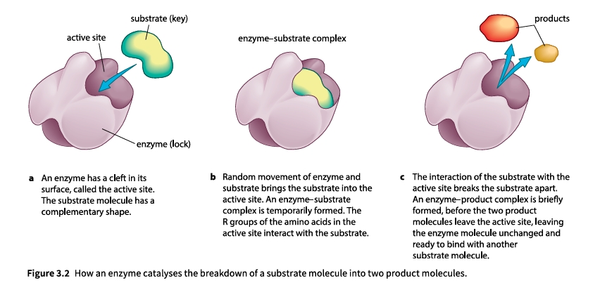
While the initial Lock and Key hypothesis suggested a completely rigid fit between the substrate and active site , modern evidence supports the Induced Fit hypothesis . The active site and substrate adjust their shape slightly upon binding to optimize catalytic interaction .
Interactive Activation Energy Diagram
Enzymes lower the activation energy (Ea) required to reach the transition state, allowing reactions to occur rapidly at normal body temperatures .
Enzyme Kinetics (Vmax and Km)
As substrate concentration [S] increases, reaction velocity (v) rises because active sites are occupied more frequently . When all active sites are saturated, the reaction reaches its maximum theoretical velocity (Vmax) .
Michaelis-Menten Constant (Km)
The Michaelis-Menten constant (Km) is the substrate concentration at which the reaction velocity is half of Vmax .
- Low Km: High affinity for substrate; reaches Vmax at lower substrate concentrations .
- High Km: Low affinity for substrate; requires higher substrate concentrations to reach saturation .
Factors Affecting Activity & Inhibition
Types of Reversible Inhibition
- Competitive Inhibition: Inhibitor resembles the substrate shape and competes directly for binding at the active site . Overcome by increasing [S] .
- Non-Competitive Inhibition: Inhibitor binds to an allosteric site elsewhere on the enzyme, distorting active site tertiary structure . Cannot be overcome by adding more substrate .
Immobilised Enzymes
Immobilisation involves trapping enzymes in inert matrices (such as calcium alginate beads) . This industrial technique allows enzymes to be reused easily and prevents product contamination .
Lactase enzyme is immobilized inside sodium alginate beads packed into a column . Milk passing through the column has its lactose hydrolyzed into glucose and galactose, yielding lactose-free milk without enzyme contamination .
Advantages of Enzyme Immobilisation
- Reusability: Enzymes are easily recovered and used in repeated industrial cycles, cutting operational costs .
- Product Purity: Products do not contain enzyme residue, eliminating downstream filtration steps .
- Enhanced Thermal & pH Stability: Matrix entrapment limits structural movement, making enzymes resistant to denaturation from heat and pH shifts .
Chapter Summary
1. Mechanism & Catalysis
Enzymes lower reaction activation energy . Substrates bind to active sites forming temporary enzyme-substrate complexes according to the induced fit model .
2. Kinetic Metrics
Vmax is maximum reaction rate at full active site saturation . Km is the substrate concentration at ½ Vmax; lower Km indicates higher enzyme affinity .
3. Rate Factors
Activity is influenced by enzyme concentration, substrate concentration, pH, and temperature . High temperatures or extreme pH denature tertiary structure .
4. Inhibition & Applications
Competitive inhibitors compete for the active site . Non-competitive inhibitors alter active site shape from afar . Immobilised enzymes in alginate beads increase stability and reusability .
Structure of Membranes
The phospholipid bilayer is visible using an electron microscope at high magnifications (≥ ×100,000). The double black line represents the hydrophilic heads of the two phospholipid layers, while the pale middle zone shows the non-polar hydrophobic interior. The total thickness of the membrane is about 7 nm on average.
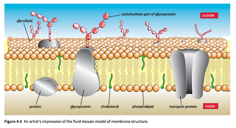
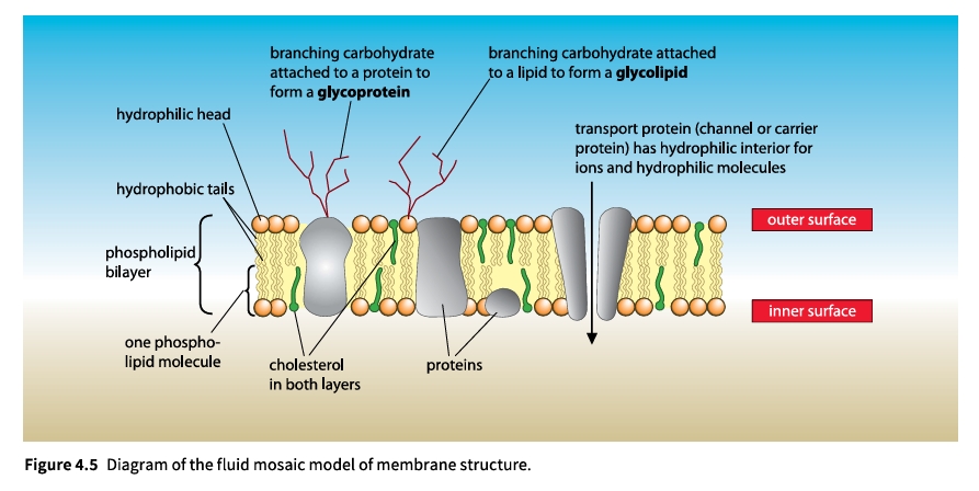
Features of the Fluid Mosaic Model (Singer & Nicolson, 1972)
Key Components & Properties
- Phospholipids: Hydrophilic heads face outward toward aqueous media; non-polar hydrophobic tails point inward. Fluidity increases with unsaturated fatty acids and shorter tail lengths.
- Intrinsic (Integral) Proteins: Embedded within the membrane. Transmembrane proteins span the entire bilayer with hydrophobic regions aligning with fatty acid tails.
- Extrinsic (Peripheral) Proteins: Bound to inner/outer surfaces.
- Glycoproteins & Glycolipids: Branching carbohydrate chains acting as cell markers and receptors.
- Cholesterol: Regulates fluidity and mechanical stability.
Interactive Phospholipid Behavior
Select an environmental configuration to render structural orientation:
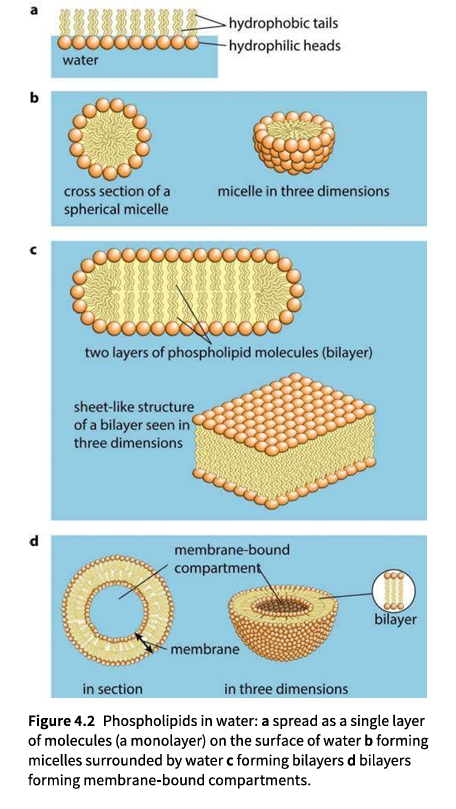 Figure 4.2: Phospholipids in water: monolayer, micelle, bilayer, and compartment.
Cell Signalling
Cell signalling is the essential biological process by which living organisms control and coordinate their bodies across cells, tissues, and systems.
Core Principles & Transduction
- Definition: Signalling is getting a message from one location to another, enabling appropriate response mechanisms.
- Transduction: Converting an original stimulus or signal into a transmitted message crossing cell surface membranes.
- Transport Distances: Short distances via local diffusion or long distances through vascular systems.
- Stimulus Diversity: Internal signals (hormones) or external factors (light, drugs).
→
1. Hydrophobic Signalling Molecules
Hydrophobic molecules, such as lipid-soluble steroid hormones (e.g., oestrogen), diffuse directly across the non-polar hydrophobic core.
- They bind directly to intracellular receptors in the cytoplasm or nucleus.
- Example: Oestrogen receptors reside within the nucleus and directly regulate gene expression.
2. Water-Soluble Signalling Molecules
Water-soluble signals cannot cross the lipid bilayer. They bind to external protein receptors embedded in the cell surface membrane.
- Specific Recognition: Receptors feature precise binding sites matching specific signalling molecules.
- Conformational Change: Alters receptor shape, transmitting the signal across the membrane.
G Proteins, Second Messengers & Signalling Cascades
A conformational change in a membrane-spanning receptor activates a membrane-bound G protein, which acts as a molecular switch.
- G Protein Mechanism: Switch mechanism involves binding to guanosine triphosphate (GTP).
- Second Messengers: Stimulate enzymes to release small, soluble molecules rapidly diffusing through cytoplasm.
- Signal Amplification: A single activated receptor produces many second messenger molecules, magnifying the signal exponentially.
Alternative Receptor Mechanisms
Besides second messengers, receptors alter cell activity through three primary mechanisms:
- Ion Channel Gating: Opening ion channels to alter membrane potential.
- Membrane-Bound Enzymes: Acting directly as an enzyme upon ligand binding.
- Intracellular Receptors: Direct control of nuclear gene expression.
Direct Cell-to-Cell Contact
Apart from secreted chemicals, direct contact between adjacent cell surface membranes serves as a vital signalling mechanism (Figure 4.8).
- Embryonic Development: Guides cell differentiation and tissue organization.
- Immune Recognition: Lymphocytes detect foreign antigens directly on target cells.
Movement of Substances Into and Out of Cells
The phospholipid bilayer forms an effective barrier against water-soluble molecules and ions. Exchange occurs via five mechanisms: diffusion, facilitated diffusion, osmosis, active transport, and bulk transport.
1. Simple Diffusion
Diffusion is the net movement of molecules or ions down a concentration gradient resulting from random motion until equilibrium is reached.
Steeper gradients result in a higher difference in molecular passing rates, increasing diffusion speed.
Higher temperatures increase kinetic energy, accelerating molecular movement.
Larger exchange surfaces allow more simultaneous crossings (e.g., microvilli, cristae).
Non-polar molecules dissolve in non-polar tails and cross rapidly. Small size helps water diffuse quickly.
Practical Investigations & Interactive Simulations
Visking tubing allows small molecules like glucose to pass through while blocking large macromolecules like starch.
Environmental stress damages membranes, causing pigment leakage via diffusion.
Tests how surface-area-to-volume ratios limit transport rates in larger structures.
2. Facilitated Diffusion
Diffusion through transport proteins providing hydrophilic pathways for large polar molecules or charged ions.
- Channel Proteins: Water-filled gated pores for charged ions.
- Carrier Proteins: Change shape to alternate binding sites between membrane sides.
Clinical Application: Cystic Fibrosis
Caused by a defect in channel proteins moving chloride ions (Cl-) out of lung cells, impairing fluid balance.
3. Osmosis
Osmosis is diffusion of water from a dilute solution to a concentrated solution across a partially permeable membrane.
- Initial State: Solution A is dilute; Solution B is concentrated. Large solute molecules bounce back.
- Net Movement: Water moves randomly, with net movement from A to B.
- Equilibrium: Solution A concentrates while B dilutes, increasing fluid volume in B.
Water Potential, Active Transport & Bulk Transport
1. Water Potential (Ψ) Fundamentals
Water potential (Ψ) measures the tendency of water to move out of a solution, dependent on solute concentration and applied pressure.
| Component | Symbol | Definition |
|---|---|---|
| Pure Water | Ψ | Pure water at atmospheric pressure has a value of 0. |
| Solute Potential | Ψs | Decreases water potential. Always 0 or negative. |
| Pressure Potential | Ψp | Physical pressure. Increases water potential. |
2. Osmotic Effects on Living Cells
- Hypotonic: Water enters, cell bursts (haemolysis).
- Isotonic: No net movement; shape constant.
- Hypertonic: Water leaves, cell shrinks (crenated).
- Pure Water: Protoplast expands against wall, becoming turgid.
- Incipient Plasmolysis: Ψp = 0, protoplast just about to separate.
- Plasmolysed: Protoplast shrinks away from the cell wall.
3. Active Transport Mechanism
Active transport moves substances against a concentration gradient using energy from ATP.
Pumps 3 Na+ OUT while moving 2 K+ IN per ATP hydrolysed, generating membrane potential.
4. Bulk Transport
Bulk uptake of solids (phagocytosis) or liquids (pinocytosis) by infolding of the cell membrane using ATP.
Bulk removal of materials via vesicle fusion with the cell surface membrane, requiring ATP.
Lesson Summary
- Fluid Mosaic Structure: Membranes consist of a phospholipid bilayer with mobile hydrophobic tails and hydrophilic heads, integrated with proteins and cholesterol.
- Component Roles: Phospholipids block polar molecules; cholesterol regulates stability; transport proteins handle passage.
- Cell Signalling: Signal transduction via receptors, G-proteins, and second messenger cascades.
- Passive Transport: Simple and facilitated diffusion move solutes down concentration gradients without ATP.
- Active & Bulk Transport: Active transport and bulk transport handle movement against gradients or in bulk using ATP.
- Osmosis & Water Potential: Water moves down a water potential (Ψ) gradient governed by solute and pressure potentials.
1. Chromosomes & Chromatin Structure
Chromosomes are thread-like structures composed of chromatin—a complex of DNA and basic histone proteins. Human cells contain 46 chromosomes. Before nuclear division occurs (during Interphase), each DNA molecule replicates. The resulting chromosome consists of two identical sister chromatids joined at a narrow region called the centromere.
Euchromatin vs. Heterochromatin
- Euchromatin: Loosely coiled, transcriptionally active DNA dominant during Interphase.
- Heterochromatin: Tightly coiled, densely staining, transcriptionally inactive DNA.
Nucleosomes
The structural unit of chromatin. Consists of 147 base pairs of DNA wrapped 1.5 times around an octamer core of 8 histone proteins. Linker DNA (53 bp) connects adjacent nucleosomes.
2. The Mitotic Cell Cycle
The cell cycle consists of three main phases: Interphase, Mitosis (M phase), and Cytokinesis.
- G₁ Phase (Gap 1): Cell grows, synthesizes RNA, enzymes, and structural proteins required for growth.
- S Phase (Synthesis): DNA replicates so each chromosome consists of two chromatids. Kinetochores begin forming.
- G₂ Phase (Gap 2): Continued growth, checking/repairing replicated DNA, and increased tubulin synthesis for spindle fibers.
- M Phase (Mitosis): Nuclear division dividing genetic material into two identical nuclei.
- Cytokinesis: Division of the cytoplasm. Constriction from edges in animal cells; cell plate formation in plant cells.
3. Telomeres & Stem Cells
Telomeres: Repeated guanine/cytosine-rich non-coding DNA sequences at chromosome ends. They prevent gene loss during DNA replication because DNA polymerase cannot copy to the extreme end of the strand. The enzyme telomerase extends telomeres in stem cells and cancer cells. Fully differentiated cells lack active telomerase, causing gradual telomere shortening and eventual cell death.
Stem Cell Potency
| Potency | Description | Examples |
|---|---|---|
| Totipotent | Can form any cell type including extraembryonic tissues (placenta). | Zygote, early blastomeres (≤16-cell stage) |
| Pluripotent | Can form all body cell types, but not extraembryonic tissues. | Embryonic stem cells |
| Multipotent | Can form a limited range of specialized cell types. | Adult bone marrow stem cells |
4. Carcinogenesis & Cancer Development
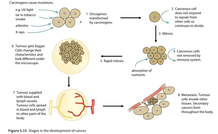
Uncontrolled mitosis caused by mutated genes (oncogenes) leads to an irregular mass of cells called a tumour. Mutagenic agents are known as carcinogens (e.g., UV, tobacco tar, asbestos, X-rays).
Metastasis: Malignant tumour cells break away, travel via blood/lymphatic vessels, and establish secondary tumours in other body tissues.
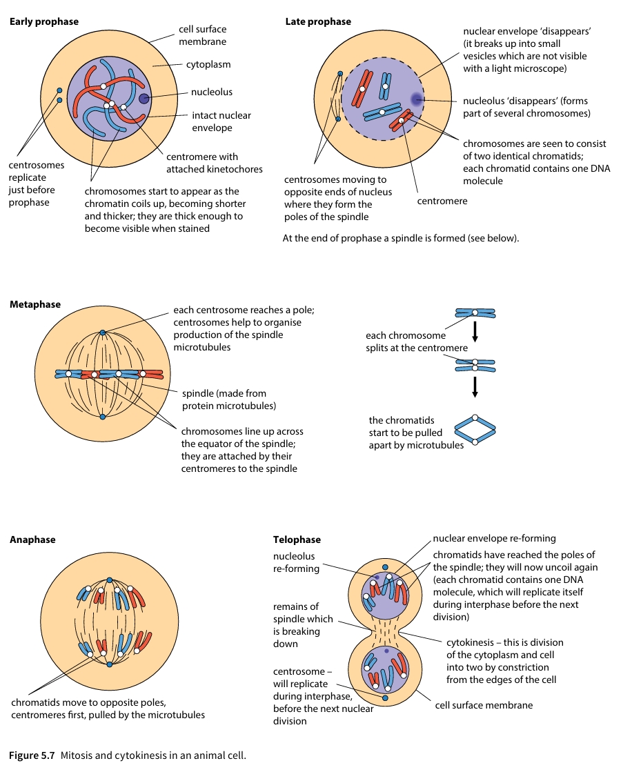
Mitosis and cytokinesis in an animal cell
Interactive Diagram Viewer
Select a diagram below to examine detailed structural models generated using responsive vector graphics:
Interactive Mitosis & Spindle Dynamics Canvas
Click the buttons below to switch stages or toggle continuous playback:
Summary & Self-Check
Comprehensive Chapter Summary
- Growth of a multicellular organism is a result of parent cells dividing to produce genetically identical daughter cells.
- During cell division, the nucleus divides first, followed by division of the whole cell. Division of a nucleus to produce two genetically identical nuclei is achieved by the process of mitosis. Mitosis is used in growth, repair, asexual reproduction and cloning of cells during an immune response.
- Although a continuous process, for convenience, mitosis can be divided into four phases: prophase, metaphase, anaphase, telophase. The period between successive nuclear and cell divisions is called interphase. During interphase chromosomes are visible only as loosely coiled material called chromatin. Chromatin consists of DNA held in position by basic proteins called histones in subunits known as nucleosomes.
- During prophase of mitosis, chromatin condenses (coils up more tightly) to form chromosomes which are easily visible in the light microscope when stained. Each chromosome consists of two identical chromatids, held together by a centromere. Each chromatid contains a single DNA molecule, formed when DNA replicates during interphase.
- The period from one cell division to the next is called the cell cycle. This has four phases: G₁ is the first growth phase after cell division; S phase is when the DNA replicates (S for synthesis); G₂ is a second growth phase; M phase (M for mitosis) is when nuclear division takes place (followed by cell division). G stands for 'gap' (between the S and M phases).
- The ends of chromosomes are capped with special regions of DNA known as telomeres. Telomeres are needed to prevent the loss of genes from the ends of chromosomes during replication of DNA.
- Many specialised cells lose the ability to divide, but certain cells known as stem cells retain this ability. Stem cells are essential for growth from zygote to adult and for cell replacement and tissue repair in the adult.
- The behaviour of chromosomes during mitosis can be observed in stained preparations of root tips, either in section or in squashes of whole root tips.
- Cancers are tumours resulting from repeated and uncontrolled mitosis. They are thought to start as the result of mutation.
Self-Assessment Challenge
Test your knowledge based on the core learning outcomes:
Q: A human cell enters mitosis. How many chromatids and kinetochores are present at metaphase?
Nucleotide & Nucleic Acid Structure
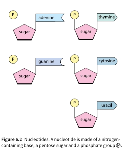
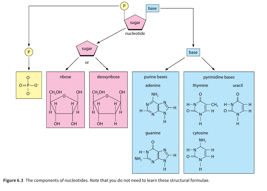
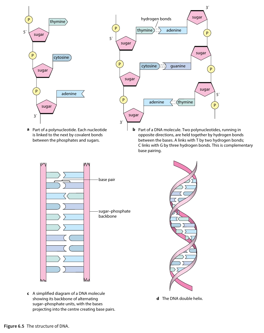
Nucleic acids (DNA and RNA) are macromolecules made of repeating units called nucleotides. Each nucleotide consists of three fundamental components:
1. Pentose Sugar
A 5-carbon sugar: Deoxyribose in DNA (lacks one oxygen atom) or Ribose in RNA.
2. Phosphate Group
An acidic, negatively charged group (PO43−) that links adjacent sugars via phosphodiester bonds.
3. Nitrogenous Base
Nitrogen-containing rings:
- Purines (double ring): Adenine (A), Guanine (G)
- Pyrimidines (single ring): Cytosine (C), Thymine (T) [DNA only], Uracil (U) [RNA only]
Key Structural Differences
DNA consists of two antiparallel polynucleotide strands running in 5′ → 3′ and 3′ → 5′ directions held together by hydrogen bonds between complementary base pairs (A = T via 2 bonds, C ≡ G via 3 bonds). RNA is single-stranded, contains ribose, and uses Uracil instead of Thymine.
Semi-Conservative DNA Replication
During S-phase of interphase, DNA replicates semi-conservatively: each original strand serves as a template to form a new daughter strand.
Mechanism Steps
- Unwinding: DNA Helicase breaks hydrogen bonds between bases, unzipping the double helix.
- Activation: Free nucleoside triphosphates (activated nucleotides) pair up with exposed bases via complementary base pairing.
- Polymerization: DNA Polymerase links adjacent nucleotides via phosphodiester bonds, releasing two inorganic phosphate groups.
- Directionality: DNA Polymerase moves in the 5′ → 3′ direction along the new strand.
BOX 6.1: Experimental Evidence for Semi-Conservative Replication
In the 1950s, three main hypotheses were proposed for how DNA replicates[cite: 2]:
1. Conservative
One completely new double helix is synthesized from the intact original molecule[cite: 2].
2. Semi-Conservative
Each newly formed DNA molecule contains one original parent strand and one newly synthesized strand.
3. Dispersive
Each new strand consists of random fragments of old and new DNA interspersed throughout the molecules[cite: 2].
The Meselson and Stahl Experiment (1958)
Matthew Meselson and Franklin Stahl tested these hypotheses using Escherichia coli (E. coli) grown in media containing isotopes of nitrogen[cite: 2]:
- Bacteria were initially cultured in a medium containing the heavy isotope nitrogen-15 (15N) until all their DNA contained heavy nitrogen[cite: 2].
- The bacteria were then transferred to a light medium containing nitrogen-14 (14N) and allowed to replicate over several generations[cite: 2].
- DNA was extracted at each generation, placed in a solution of caesium chloride, and separated by density using high-speed centrifugation[cite: 2].
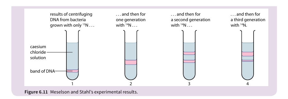
1: DNA grown entirely in 15N forms a single heavy band near the bottom of the tube[cite: 2].
2: Generation 1 in 14N produces a single band of intermediate density (15N-14N hybrid DNA), ruling out conservative replication[cite: 2].
3: Generation 2 in 14N produces two equal bands: one intermediate band and one light band (14N-14N), ruling out dispersive replication and proving semi-conservative replication[cite: 2].
4: Generation 3 in 14N shows an increased proportion of light DNA relative to intermediate DNA[cite: 2].
The Triplet Code & Gene Mutations
The genetic sequence is read as a continuous series of 3-base units called codons (triplets).
Triplet Code Features
- Universal: Same codons specify identical amino acids across virtually all organisms[cite: 2].
- Degenerate: 64 possible triplets (43) code for 20 amino acids; multiple triplets code for the same amino acid[cite: 2].
- Non-overlapping: Each base is part of only one triplet[cite: 2].
Sickle Cell Anaemia Case Study
A single base substitution mutation alters the β-globin polypeptide chain:
Normal (HbA): DNA triplet CTT → Glutamic acid.
Sickle (HbS): DNA triplet CAT → Valine.
Effect: Non-polar valine alters hemoglobin solubility, causing cell sickling and low oxygen transport capabilities.
Transcription and Translation
1. Transcription (Nucleus)
RNA Polymerase binds to the promoter, unwinds DNA, and aligns free RNA nucleotides along the template strand (3′ → 5′). It links nucleotides to synthesize a complementary single-stranded mRNA molecule, stopping at a terminator sequence.
2. Translation (Ribosome in Cytoplasm)
mRNA binds to the small ribosomal subunit, exposing two codons at a time. tRNA molecules with matching anticodons deliver specific amino acids. Peptidyl transferase forms peptide bonds between neighboring amino acids until a STOP codon is reached.
Interactive Transcription & Translation Simulator
Select a DNA template strand to transcribe into mRNA and translate into an amino acid chain.
Summary
- DNA and RNA are polynucleotides, made up of long chains of nucleotides . A nucleotide contains a pentose sugar, a phosphate group and a nitrogen-containing base . In RNA, the sugar is ribose; in DNA, it is deoxyribose .
- A DNA molecule consists of two polynucleotide chains, linked by hydrogen bonds between bases . In DNA there are four bases – adenine always pairs with thymine, and cytosine always pairs with guanine .
- RNA, which comes in several different forms, has only one polynucleotide chain, although this may be twisted back on itself, as in tRNA . In RNA, the base thymine is replaced by uracil .
- DNA molecules replicate during interphase by semi-conservative replication . In DNA replication, the hydrogen bonds between the bases break, allowing free nucleotides to fall into position opposite their complementary ones on each strand of the original DNA molecule . Adjacent nucleotides are then linked, through their phosphates and sugars, to form new strands . Two complete new molecules are thus formed from one old one, each new molecule containing one old strand and one new strand .
- The sequence of nucleotide bases on a DNA molecule codes for the sequence of amino acids in a polypeptide . Each amino acid is coded for by three bases . A length of DNA coding for just one polypeptide is a gene . A change in the nucleotide sequence of DNA is a mutation, producing a new allele of the gene .
- The DNA sequences for the HbA (normal) and HbS (sickle cell) alleles of the gene for the β-globin polypeptide differ by only one base . The triplet CTT in HbA is replaced by CAT in HbS, swapping the amino acid from glutamic acid to valine . This single difference in the polypeptide results in sickle cell anaemia in individuals with two HbS alleles .
- During protein synthesis, a complementary copy of the base sequence on a gene is made, by building a molecule of messenger RNA (mRNA) against one DNA strand . This stage is called transcription . After transcription, the next stage is called translation . During translation the mRNA moves to a ribosome in the cytoplasm .
- Transfer RNA (tRNA) molecules with complementary triplets of bases temporarily pair with base triplets on the mRNA, bringing appropriate amino acids . When two amino acids are held side by side, a peptide bond forms between them . The ribosome moves along the mRNA molecule, so that appropriate amino acids are gradually linked together, following the sequence laid down by the base sequence on the mRNA .
Plant Transport Needs & Tissue Anatomy
Unlike animals, plants do not possess a central mechanical pump or a rapid gaseous transport circulation system. Photosynthetic gases (CO₂ and O₂) move independently via passive diffusion through connected intercellular air spaces.
- Absorbed to Required: Water and inorganic mineral ions absorbed by roots must move upward to photosynthetic stems and leaves.
- Produced to Required: Assimilates (e.g., sucrose) manufactured in leaves during photosynthesis must be transported to metabolic sinks (roots, buds, tubers).
- Storage Movements: Sugars are transported to storage organs (e.g., roots/tubers) to be synthesized into insoluble starch.
Overall Water Movement Through a Plant
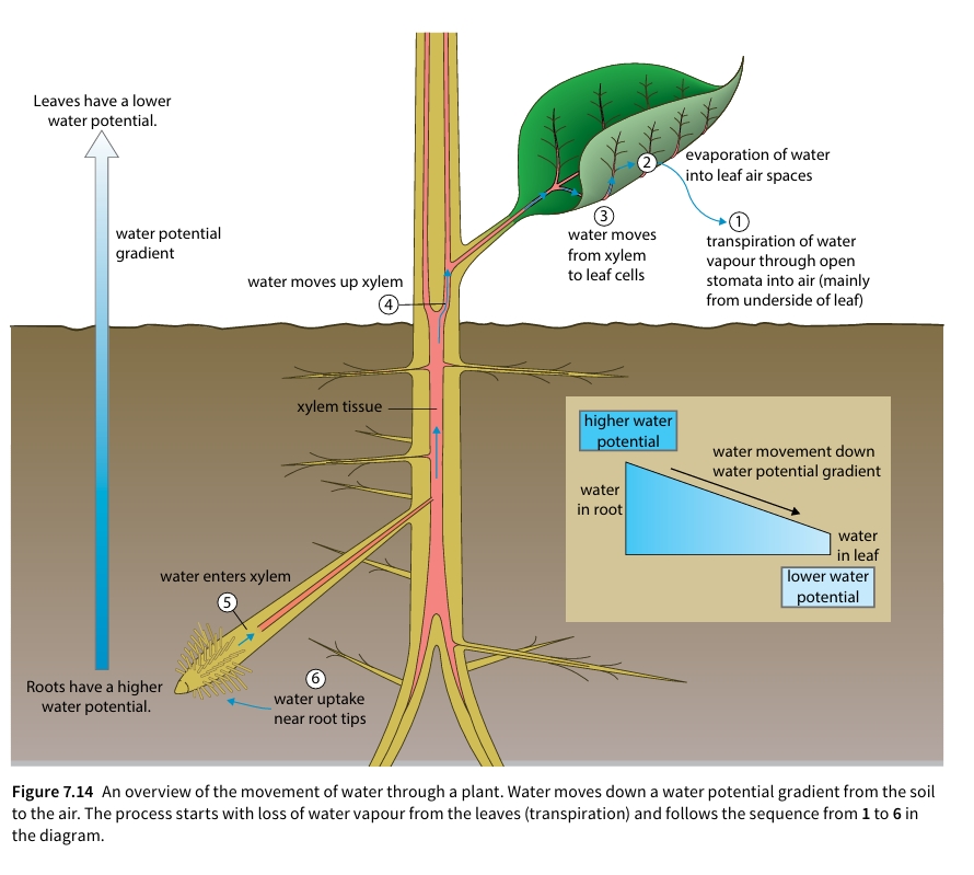
Water moves down a continuous water potential gradient from regions of higher water potential ($\Psi$) in the soil through the root tips and xylem tissue to the lower water potential of ambient air spaces in leaves.
Organ Cross-Sections (Dicotyledonous Structures)
Vascular tissues (xylem and phloem) are arranged distinctly in dicot stems, roots, and leaves to handle specialized structural stresses:
Tissue Structure Summary
| Tissue Type | Cell Types / Structure | Primary Function |
|---|---|---|
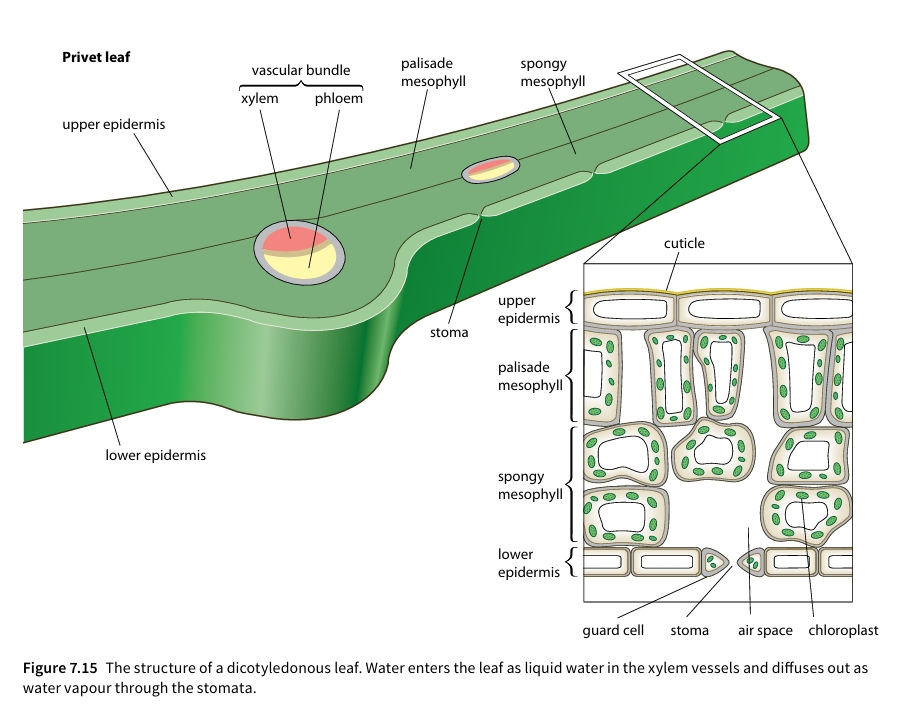
| Epidermis | Single layer, waxy cuticle, contains stomata/guard cells | Protects against water loss and pathogen entry |
| Parenchyma | Thin primary cellulose walls, unspecialized, air spaces | Packing tissue, starch storage, turgor support |
| Collenchyma | Living cells with extra cellulose thickening at corners | Flexible mechanical support in stems & leaf midribs |
| Endodermis | Inner cell boundary surrounding vascular cylinder; Casparian strip | Selectively controls ion entry into root xylem |
| Sclerenchyma | Dead cells with thick, lignified secondary walls | Rigid mechanical support (fibres in vascular bundles) |
Detailed Dicotyledonous Leaf Anatomy
Xylem Vessel Structure & Pathway of Water
Water moves passively from regions of higher water potential ($\Psi$) in the soil to lower water potential in the air. Xylem vessels consist of dead, hollow, elongated vessel elements linked end-to-end. End walls break down completely to form continuous drainpipe-like tubes. Secondary cell walls are reinforced with lignin to withstand negative hydrostatic pressure (tension) without collapsing. Unlignified gaps called pits permit lateral water movement between adjacent vessels.
Root Tissue Pathways (Apoplast vs. Symplast)
Water absorbed by root hairs crosses the root cortex via two primary pathways:
- Apoplast Pathway: Water movement through non-living cellulose cell walls and intercellular spaces by capillary movement. Blocked at the endodermis by the waterproof Casparian strip (suberin).
- Symplast Pathway: Water moves through cytoplasm and living cell interiors linked by plasmodesmata via osmosis.
Transpiration & Potometer Simulation
Transpiration is the loss of water vapour from a plant to its environment by diffusion down a water potential gradient, predominantly taking place through leaf stomata.
- Solar radiation causes liquid water to evaporate from wet mesophyll cell walls into internal sub-stomatal air spaces.
- Water vapour diffuses out through open stomata into drier surrounding air down a water potential gradient.
- Transpiration lowers hydrostatic pressure at the top of xylem vessels, creating tension.
- Water is pulled up xylem in a continuous mass-flow column, facilitated by cohesion (H-bonding between water molecules) and adhesion (attraction to lignified walls).
Interactive Potometer Simulation
A potometer estimates transpiration rate by measuring the rate of water uptake by a cut plant shoot. Adjust environmental variables to observe capillary meniscus movement.
Xerophytic Leaf Adaptations
Xerophytes are specialized plants adapted to habitats with limited water supply or high potential transpiration demand. They possess anatomical structural modifications to restrict transpiration loss.
Xerophytic Structural Features
| Adaptation | Mechanism for Water Conservation | Example Plant |
|---|---|---|
| Rolled Leaves | Traps humid air inside roll; reduces exposure of stomata to dry air currents. | Marram grass (Ammophila) |
| Sunken Stomata | Pits trap a boundary layer of moist air, reducing water potential gradient. | Sitka spruce, Marram grass |
| Epidermal Hairs (Trichomes) | Physical barrier trapping humid air layer, reflecting light. | Phlomis italica, Marram grass |
| Thick Waxy Cuticle | Impenetrable hydrophobic cutin layer minimizes cuticular evaporation. | Cactus (Opuntia), Conifers |
| Needle/Spine Leaves | Drastically reduces surface area-to-volume ratio. | Spruce, Cacti (Opuntia) |
Phloem Structure & Mass Flow Translocation
Translocation is the energy-requiring transport of soluble organic assimilates (predominantly sucrose and amino acids) through phloem tissue from sources (e.g. photosynthesizing mature leaves) to sinks (e.g. growing root tips, developing seeds, tubers).
Phloem Sieve Tube & Companion Cell Structure
- Sieve Tube Elements: Living cells joined end-to-end. Lacks nuclei, microfilaments, or ribosomes to reduce resistance to fluid flow. End walls form perforated sieve plates.
- Companion Cells: Metabolically active helper cells connected to sieve tube elements via high-density plasmodesmata. Contain abundant mitochondria and rough ER to generate ATP required for active loading.
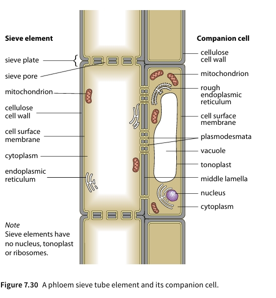
Active Sucrose Loading Mechanism
- H⁺ Ion Pumping: Companion cells use ATP to actively pump $H^+$ ions across their membrane into the apoplastic cell wall space.
- Co-Transport: $H^+$ ions diffuse back into companion cells down their concentration gradient through $H^+$/sucrose co-transporter carrier proteins, moving sucrose against its concentration gradient.
- Symplastic Entry: Sucrose moves from companion cells into sieve tube elements through interconnecting plasmodesmata.
- Osmotic Hydrostatic Pressure: Accumulation of sucrose dramatically lowers $\Psi$ inside the sieve element. Water enters from xylem by osmosis, generating a high hydrostatic pressure that drives mass flow toward sinks.
Chapter Summary
1. Transport Requirements
- Multicellular organisms with small surface area : volume ratios need specialized transport systems .
- Flowering plants do not have compact bodies like animals; they spread and branch above and below ground to obtain carbon dioxide, light energy, water, and mineral ions .
- Plants do not need systems for transporting carbon dioxide or oxygen—diffusion is sufficient .
2. Water Movement & Xylem
- Water and mineral salts are transported through a plant in xylem vessels .
- Movement through the plant is a passive process down a water potential gradient from soil to air .
- Energy comes from the Sun, causing water evaporation from wet mesophyll cell walls in leaves .
- Water vapour diffuses out through stomata via transpiration, setting up the water potential gradient throughout the plant .
- Water enters root hairs by osmosis, crosses the root via cytoplasm (symplastic pathway) or cell walls (apoplastic pathway), and enters dead, empty xylem vessels .
- Water moves across the leaf by both symplast and apoplast pathways .
- Xylem sap moves up vessels by mass flow due to pressure differences from leaf transpiration; root pressure can also contribute .
- Xylem transport is strictly unidirectional (from roots to the rest of the plant) .
3. Transpiration & Potometer
- Transpiration is an inevitable consequence of gaseous exchange needed for CO₂ and O₂ exchange through stomata .
- Transpiration rate is affected by environmental factors: temperature, light intensity, wind speed, and humidity .
- Measuring transpiration rate directly is difficult, but water uptake can be measured using a potometer .
4. Xerophytes
- Xerophytes are adapted to places where conditions cause high transpiration rates and soil water is in short supply .
- They have evolved structural adaptations that help reduce the rate of water vapour loss from leaves .
5. Phloem & Translocation
- Translocation of organic solutes (e.g., sucrose) occurs through living phloem sieve tubes .
- Phloem sap moves by mass flow from source to sink .
- Sucrose is produced at the source (e.g., photosynthesising leaves) and used at the sink (e.g., a flower or storage organ) .
- Active loading of sucrose at the source causes water entry by osmosis, creating high hydrostatic pressure in sieve tubes .
- Phloem sap can move in different directions in different sieve tubes .
6. Structural Adaptations
- Both xylem vessels and phloem sieve tubes show unique structural features and distributions adapted specifically to their transport roles .
Comparative Overview: Xylem vs. Phloem
| Feature | Xylem Vessels | Phloem Sieve Tubes |
|---|---|---|
| Living Status | Dead at maturity (empty hollow tubes) | Living cells |
| Substances Carried | Water and mineral salts / ions | Organic solutes (e.g. sucrose) |
| Direction of Flow | Unidirectional (roots to rest of plant) | Multidirectional (different directions in different tubes) |
| Transport Mechanism | Passive mass flow driven by transpiration & root pressure | Mass flow driven by hydrostatic pressure from active sucrose loading |
1. Mammalian Circulatory System & Blood Vessels
Mammals possess a closed double circulatory system. Blood remains inside blood vessels and passes through the heart twice during one complete circuit around the body:
- Pulmonary Circulation: Deoxygenated blood is pumped from the right ventricle to the lungs via pulmonary arteries and returned oxygenated to the left atrium via pulmonary veins.
- Systemic Circulation: Oxygenated blood is pumped from the left ventricle through the aorta to the rest of the body tissues, returning deoxygenated to the right atrium via the vena cava.
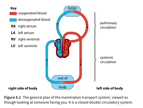
Figure 8.2: The general plan of the mammalian transport system, viewed as though looking at someone facing you. It is a closed double circulatory system.
Comparison of Blood Vessels
Blood vessel walls (except capillaries) consist of three main layers (tunicae): tunica intima (endothelium), tunica media (smooth muscle and elastic tissue), and tunica externa (collagen fibers).
| Vessel Type | Lumen Size | Wall Thickness & Composition | Blood Pressure | Function & Key Adaptations |
|---|---|---|---|---|
| Artery | Narrow | Thick; heavy tunica media rich in elastic fibers & smooth muscle. | High (~120 to 80 mmHg) | Withstands high pressure; elastic recoil evens out pulsed blood flow. |
| Arteriole | Variable | Contains higher proportion of smooth muscle than elastic tissue. | Drops (85 to 35 mmHg) | Vasoconstriction/vasodilation controls local blood distribution & lowers pressure. |
| Capillary | Extremely narrow (~7 μm) | Single layer of endothelial cells (~1 μm thick). | Low (35 to 10 mmHg) | Exchanges nutrients/gases; gaps allow tissue fluid formation. |
| Vein | Wide | Thin; small amount of muscle/elastic tissue; presence of semilunar valves. | Very Low (<5 mmHg) | Returns blood to heart; valves prevent backflow under low pressure. |
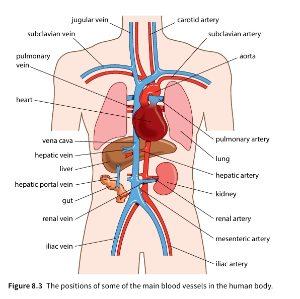
Figure 8.3: The positions of some of the main blood vessels in the human body.
2. Blood Plasma, Tissue Fluid, and Lymph
As blood flows through capillaries, hydrostatic pressure forces fluid out into surrounding intercellular spaces.
- Arterial End: Hydrostatic pressure (~4.7 kPa) exceeds osmotic pressure pulling water back in (~3.3 kPa). Fluid, glucose, amino acids, and dissolved ions are filtered out through endothelial gaps.
- Venous End: Hydrostatic pressure drops (~1.3 kPa) below osmotic pressure. Net movement of water occurs back into capillaries by osmosis.
- Large plasma proteins (e.g., albumin) and blood cells remain inside capillaries as they cannot pass through capillary pores.
The Lymphatic System
About 10% of leaked fluid does not return to capillaries and is drained by blind-ending lymphatic vessels as lymph.
- Composition: Virtually identical to tissue fluid, but in specific areas (e.g., small intestine lacteals) it contains digested lipids.
- Drainage Path: Lymphatic vessels contain valves to enforce one-way flow, passing through lymph nodes (where white blood cells remove pathogens) before emptying back into the blood via the subclavian veins.
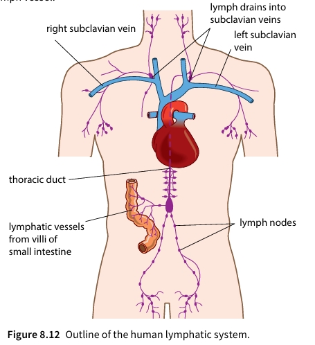
Figure 8.12: Outline of the human lymphatic system.
3. Blood Components & Gas Transport Dynamics
Cellular Components of Blood
- Erythrocytes (Red Blood Cells): Biconcave discs (~7 μm diameter) lacking a nucleus, ER, or mitochondria. Packed with haemoglobin for maximum oxygen-carrying capacity.
- Leucocytes (White Blood Cells): Possess nuclei; involved in defense. Divided into Phagocytes (Neutrophils with lobed nuclei and Monocytes) and Lymphocytes (large round nuclei producing antibodies or cellular immunity).
- Platelets: Cell fragments involved in blood clotting.
Haemoglobin (Hb) is a tetrameric protein that binds reversibly with up to 4 oxygen molecules (Hb + 4O2 ⇌ HbO8). Binding of the first O2 alters protein tertiary structure, making subsequent bindings easier (cooperative binding), producing a sigmoidal (S-shaped) dissociation curve.
The Bohr Shift: In actively respiring tissues, high CO2 production lowers blood pH via carbonic acid dissociation (CO2 + H2O carbonic anhydrase⟶ H2CO3 → H+ + HCO3-). H+ ions bind to haemoglobin forming haemoglobinic acid (HHb), reducing Hb affinity for oxygen and shifting the curve to the right, causing increased oxygen release.
Figure 3.1: Oxygen Dissociation Curve and the Bohr Shift.
4. Heart Structure, Cardiac Cycle, & Myogenic Control
Structure of the Heart
The mammalian heart has 4 chambers: two thin-walled atria (receive blood) and two thick-walled ventricles (pump blood out). The muscular septum completely separates left and right sides. The left ventricle wall is substantially thicker than the right ventricle wall because it must pump blood through high resistance in the systemic circulation.
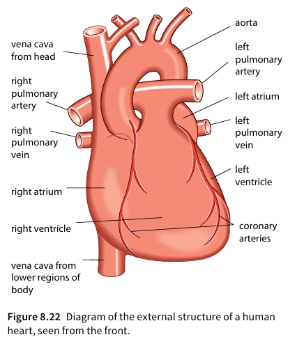
Figure 8.22: Diagram of the external structure of a human heart, seen from the front.
The Cardiac Cycle Sequence
- Atrial Systole: Atria contract; blood is forced through open atrioventricular (AV) valves (tricuspid/bicuspid) into ventricles.
- Ventricular Systole: Ventricles contract; ventricular pressure exceeds atrial pressure, snapping AV valves shut (first heart sound). Semilunar (SL) valves open, forcing blood into aorta and pulmonary artery.
- Ventricular Diastole: Muscle relaxes; ventricular pressure drops below arterial pressure, causing SL valves to snap shut (second heart sound). Blood flows passively into atria from veins.
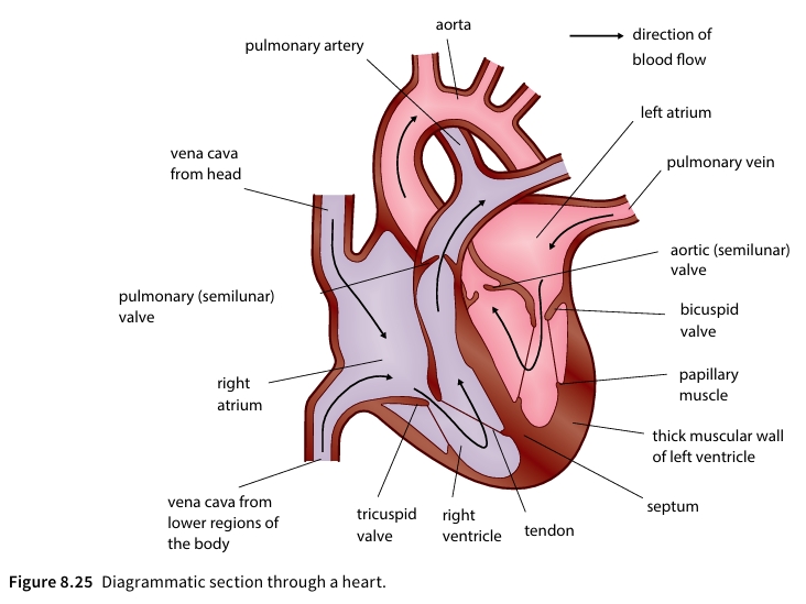
Figure 8.25: Diagrammatic section through a heart.
The heart beat originates internally without external nervous impulses:
SAN (Sinoatrial Node / Pacemaker) → Wave across Atria → AVN (Atrioventricular Node - 0.1s delay) → Bundle of His / Purkyne Tissue → Ventricles contract from Apex upwards.
5. Interactive Cardiac Conduction & Pressure Simulator
Observe real-time cardiac muscle contraction, electrical impulse conduction, and associated pressure variations during the cardiac cycle.
6. Lesson Summary
Key Takeaways
- Double Circulation: Separate pulmonary and systemic loops maintain high metabolic blood pressure to body tissues.
- Vessel Specialization: Arteries absorb high pressure pulses via elastic recoil; Arterioles regulate flow; Capillaries enable rapid diffusion; Veins store low-pressure blood with valves preventing backflow.
- Fluid Exchange: Hydrostatic pressure pushes fluid out at arterial ends; osmotic gradients pull water back at venous ends. Excess tissue fluid drains into lymphatics.
- Gas Transport: Haemoglobin displays cooperative binding. High CO2 concentrations lower blood pH, driving the Bohr shift to release O2 at active tissues.
- Myogenic Heart Cycle: The SAN initiates contraction. A brief delay at the AVN ensures full atrial emptying before Purkyne fibers trigger ventricular contraction from the apex upward.
Chapter 9: Gas Exchange and Smoking
The human gas exchange system links the circulatory system with the atmosphere. It is adapted to clean and warm incoming air, maximize surface area for diffusion, minimize diffusion distance, and maintain steep concentration gradients.
The Airway System & Structure
Air passes through a branching system of airways starting from the trachea, moving into the bronchi, bronchioles, and finally terminating in the alveoli.
| Airway | Number | Approx. Diameter | Cartilage | Goblet Cells | Smooth Muscle | Cilia |
|---|---|---|---|---|---|---|
| Trachea | 1 | 1.8 cm | Yes | Yes | Yes | Yes |
| Bronchus | 2 | 1.2 cm | Yes | Yes | Yes | Yes |
| Terminal Bronchiole | 48,000 | 1.0 mm | No | No | Yes | Yes |
| Respiratory Bronchiole | 300,000 | 0.5 mm | No | No | No | A few |
| Alveolar Duct | 9 × 10⁶ | 400 µm | No | No | No | No |
| Alveoli | 3 × 10⁹ | 250 µm | No | No | No | No |
Key Tissues and Defence Mechanisms
- Cartilage: C-shaped rings in the trachea and irregular blocks in the bronchi keep airways open and reduce air resistance.
- Goblet Cells & Mucus: Produce sticky mucus containing mucin glycoproteins to trap dust, pollen, bacteria, and viruses.
- Ciliated Epithelium: Beats continually to move trapped particles upward towards the larynx at ~1 cm/min to be swallowed and destroyed by stomach acid.
Structure of the Human Lungs
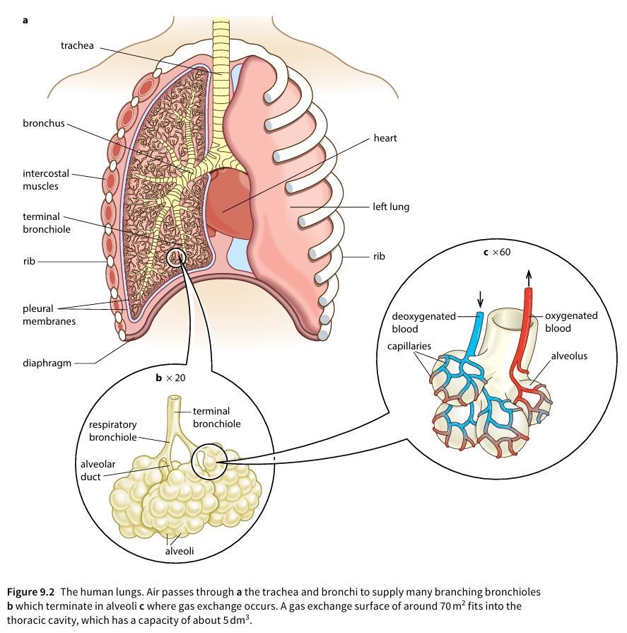
Figure 9.2 outlines the structural organization of the human respiratory system, showing how air is channeled from the upper airways down to the microscopic gas exchange surfaces.
Key Anatomical Features
- Thoracic Cavity & Pleural Membranes: The lungs sit in an airtight space enclosed by pleural membranes containing fluid for frictionless movement during ventilation.
- Branching Airways (a & b): Trachea splits into two bronchi, branching extensively into terminal and respiratory bronchioles.
- Alveolar Clusters (c): Surrounded by dense capillary networks where deoxygenated blood is converted to oxygenated blood.
Mechanisms of Gas Exchange
The alveoli provide a massive surface area of around $70\text{ m}^2$ in an adult human packed inside a thoracic cavity of about $5\text{ dm}^3$.
Adaptations of Alveoli
- Thin Walls: Single layer of squamous epithelial cells (~0.5 µm thick).
- Capillary Network: Closely pressed microvessels maintain short diffusion pathways.
- Concentration Gradients: Maintained continuously by constant blood flow and ventilation.
- Elastic Fibres: Stretch during inspiration and recoil during expiration to force air out effectively.
Diffusion Dynamics
Oxygen moves down its concentration gradient from alveolar air into the deoxygenated blood capillaries. Conversely, carbon dioxide diffuses from the blood into the alveolar air space to be exhaled.
Interactive Lung Ventilation & Gas Flow Simulation
Use the controls below to simulate normal breathing versus restricted airways caused by smoking or chronic bronchitis.
Smoking, COPD, and Cardiovascular Impacts
Tobacco smoke contains over 4,000 chemicals, many of which are toxic. The primary harmful agents are Tar, Carbon Monoxide, and Nicotine.
Chronic Obstructive Pulmonary Disease (COPD)
- Chronic Bronchitis: Tar enlarges goblet cells, overproduces mucus, and destroys cilia. Bacteria accumulate, leading to smoker's cough and scarred, narrowed airways.
- Emphysema: Phagocytes release elastase to break down alveolar walls. Elastin is destroyed, preventing proper recoil. Alveoli burst, drastically reducing surface area.
Cardiovascular Effects
- Nicotine: Stimulates nervous system release of adrenaline, increasing heart rate and blood pressure while restricting peripheral blood flow. Highly addictive via dopamine pathways.
- Carbon Monoxide: Binds irreversibly with hemoglobin to form carboxyhaemoglobin, reducing oxygen-carrying capacity by 5–10%.
Chapter Summary
- Multicellular organisms often have surfaces that are specialised to allow exchange of gases to take place between their bodies and the environment. Alveoli in the lungs form the gas exchange surface in mammals.
- In the human lungs, air passes down the trachea and through a branching system of airways to reach the alveoli. The airways are lined by a ciliated epithelium with mucus-secreting goblet cells. The epithelium protects the alveoli by moving a carpet of mucus towards the throat, where it can be swallowed. There are C-shaped rings of cartilage in the trachea and irregularly shaped blocks of cartilage in the bronchi to keep the large airways open and so reduce resistance to the flow of air. Smooth muscle in the airways contracts and relaxes to adjust the diameter of the airways.
- The alveoli are lined by a squamous epithelium that gives a short diffusion distance for the exchange of oxygen and carbon dioxide. The alveoli are well supplied with blood by the many capillaries surrounding the gas exchange surface. The constant flow of blood and the continuous ventilation of the lungs maintain concentration gradients between blood and air for oxygen and carbon dioxide. Recoil of the elastic fibres surrounding the alveoli helps to move air out during expiration.
- Damage to the airways and alveoli occurs in chronic obstructive pulmonary disease (COPD). In chronic bronchitis, the airways are obstructed by mucus and infection; in emphysema, alveoli are destroyed, reducing the surface area for gas exchange. Some of the signs and symptoms of COPD are breathlessness, wheezing and constant coughing.
- Tobacco smoke contains tar, carbon monoxide and nicotine. Tar settles on the epithelium lining the bronchi and bronchioles and stimulates inflammation, an increase in the secretion of mucus and an accumulation of phagocytes from the blood. Tar contains carcinogens, which cause changes in DNA in bronchial epithelial cells, often leading to the development of a bronchial carcinoma. This is lung cancer. Two of the symptoms of lung cancer are coughing up blood and chest pains.
- Carbon monoxide combines irreversibly with haemoglobin, reducing the oxygen-carrying capacity of the blood. Nicotine stimulates the nervous system, increasing heart rate and blood pressure, and stimulating vasoconstriction, which reduces blood flow to the extremities.
- Smoking damages the cardiovascular system, increasing and increasing the risk of coronary heart disease and stroke.